August 19, 2006
Of rats and (wo)men
I've recently read a couple of recent books on the science of sex differences: Leonard Sax's "Why Gender Matters: What Parents and Teachers Need to Know about the Emerging Science of Sex Differences", and Louan Brizendine's "The Female Brain". As I explained in previous posts, I was disappointed and even shocked at these authors' cavalier treatment of issues in speech, language and communication ("Are men emotional children?"; "Neuroscience in the service of sexual stereotypes"; "Sex and speaking rate"; "Sex-linked lexical budgets"). And as I've read more, I've learned that at least some of the core neuroscience in both books is equally shaky. More precisely, both authors erect some robust and convincing rhetorical structures on some very weak scientific foundations.
That's not to say that every piece of science that these books present is faulty -- I've only checked a few things. But what I've found, in the cases where I've checked, suggests that you'd be wise not to accept anything from such sources without looking into it carefully for yourself. There's a spectacular example in chapter 2 of Sax's book, under the heading "The Eye of the Beholder" (p. 18-29).
Sax starts by telling us a bit about the retina -- rods and cones, P and M ganglion cells -- and gives this functional account of the ganglion cells:
P cells and M cells have very different jobs. M cells are ... essentially simple motion detectors. ... You can think of the M cells as being wired to answer the questions "Where is it now and where is it going?" ... You can think of P cells as answering the question "What is it?" P cells compile information about texture and color; M cells compile information about movement and direction.
The P cells send information via their own special division of the thalamus to a particular region of the cerebral cortex that appears to be specialized for analysis of texture and color. The M cells sen their information via a separate pathway to a different region of the cerebral cortex, a region that is specialized for analysis of spatial relationships and object motion. And guess what? Every step in each pathway, from the retina to the cerebral cortex, is different in females and males.23
[emphasis original]
Cool. You can see where he's going with this: women's eyes and brains are specialized for a certain set of tasks, men's eyes and brains for a very different set of tasks. But if you follow his end-note 23 to its reference, you find: Tamas Horvath and K.C. Wikler, "Aromatase in Developing Sensory Systems of the Rat Brain," Journal of Neuroendocrinology, 11:77-84, 1999. So we might have expressed that italicized phrase differently: "Every step in each pathway, from the retina to the cerebral cortex, is different in female rats and male rats." Except, we're all mammals, right? with the same basic hormones and hormone responses? Sort of. Hold that thought, because now the emerging sexual science gets really hot:
The real surprises have come from microscopic analyses of the eye performed in the past five years. Using recently developed techniques, scientists have found that the human retina is full of receptors for sex hormones.24 Anatomist Edwin Lephart and his associates have found that the male retina is substantially thicker than the female retina.25 That's because the male retina has mostly the larger, thicker M cells while the female retina has predominantly the smaller, thinner P ganglion cells.
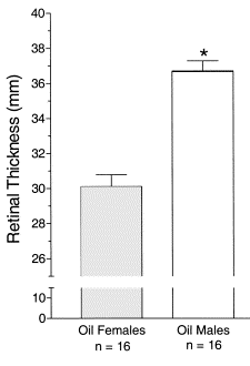
We're not talking about small differences between the sexes, with lots of overlap. We're talking about large differences between the sexes, with no overlap at all. Every male animal had a thicker retina than any female retina, due to the males having more M cells (see the accompanying graph).
That's some serious differentiation, alright, and Sax shows us the graph to prove it:

(This is not exactly Sax's figure, since I took it from his source rather than from his book, but it's the same data.)
Pretty impressive, right? I was completely blown away when I saw it, I'll confess. The only thing is, if we follow up that end-note 25, we find: David Salyer, Trent D. Lund, Donovan E. Fleming, Edwin Lephart, and Tamas L. Horvath, "Sexual dimorphism and aromatase in the rat retina", Developmental Brain Research, 126:131-136, 2001.
Rats, foiled again! (Or maybe that should be "Foiled, rats again!")
[Just for completeness, the actual figure from the Salyer et al. paper is shown and explained at the end of this post. You'll also learn there what "Oil Females" means.]
But Sax is not just a journalist or a politician -- he has an M.D. and a Ph.D, so surely he wouldn't use this figure as part of an argument about how boys and girls see the world in completely different ways, if humans weren't the same as rats in this respect. After all, humans have the same sex hormones that rats do, and the same sort of eyes and overall visual neurology, and scientists use rats all the time as model organisms for learning about human physiology. Still, it would be nice to see some data about retinal thickness in our own species, so I hit up Google Scholar with "retina thickness male female", and in about five minutes I had an answer.
Yoshikatsu Wakitani et al. , "Macular thickness measurements in healthy subjects with different axial lengths using optical coherence tomography", Retina 23(2) April 2003.
Purpose: To evaluate the retinal thickness of the macula in healthy subjects with different axial lengths.
Methods: Included were 203 healthy subjects (116 males and 87 females). The axial length of the eyes ranged from 22.68 to 30.22 mm. Four optical coherence tomograms were obtained in a radial spoke pattern centered on the central fovea. Retinal thickness was calculated from the inner and outer retinal boundaries. The average retinal thickness in three circular areas surrounding the central fovea (350, 1,850, and 2,850 [mu]m in diameter) was determined.
[The "macula" is an area of the retina around the fovea, where vision is most acute.]
And indeed, Wakitani et al. did find a significant mean difference in retinal thickness between human males and females, as this table shows:

But wait a minute -- we're supposed to be seeing completely separable distributions for males and females here, with "no overlap at all", so that "every male ... had a thicker retina than any female". However, in this table, the differences between the male and female means are only about a third to a half a standard deviation: in area A it's 9 μm difference vs. a 20-21 μm standard deviation; in area B it's a 7 μm difference vs. an 16-18 μm s.d.; in area C it's a 6 μm difference vs. a 13-15 μm s.d. (Note that these differences are also small relative to the means -- about a 4% relative difference, which is significantly less than other mean differences in linear dimensions between men and women, such as the 8% difference in height; and much less than mean differences in measurements where there is really a significant amount of sexual dimorphism in humans, such as the 60% difference between men and women in vocal cord length.)
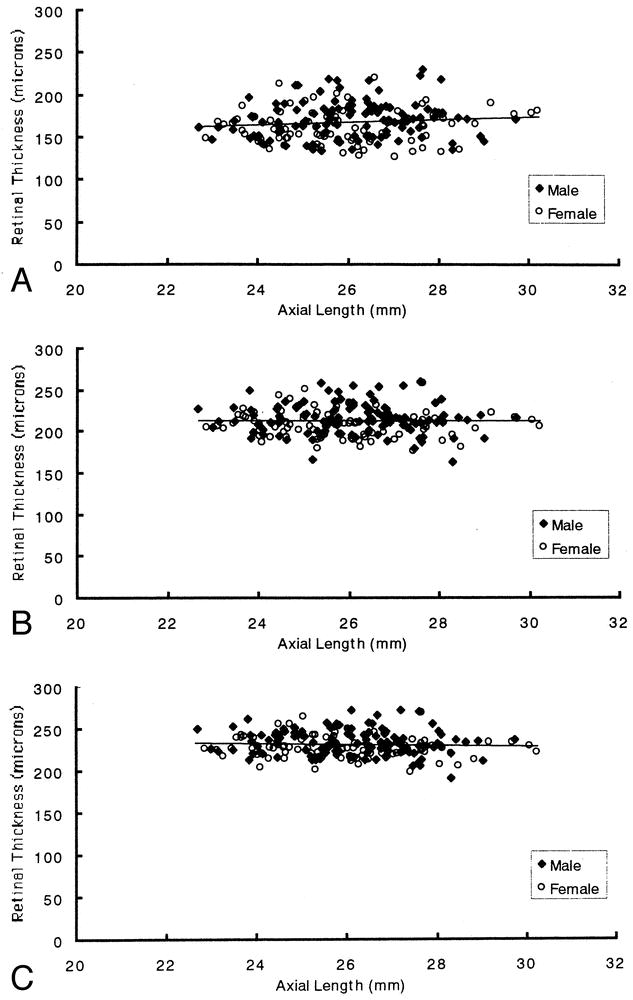
If we want a more visceral sense of what this means, Wakitani et al. provide some scatter plots:

Fig. 4. Relationship between axial length (mm) and retinal thickness (µm). A, Area A (350 µm in diameter, including the foveola). B, Area B (1,850 µm in diameter, including the fovea). C, Area C (2,850 µm in diameter, including the parafovea). Linear regression analysis that included both male and female data indicated no statistically significant change in retinal thickness with increasing axial length of the eye (P = 0.10 for area A, P = 0.39 for area B, and P = 0.12 for area C).
From: WAKITANI: Retina, Volume 23(2).April 2003.177-182
Not exactly "no overlap at all", would you say?
Sax connects these "non-overlapping" retinal differences with lots of other facts about sex differences (or perhaps I should say "facts", subject to tracking down the rest of his references): women "interpret facial expressions better"; "girls draw nouns, boys draw verbs"; "females and males use fundamentally different strategies" for "geometry and navigation"; at ages when "little children ... had not a clue which gender they belonged to", "nine-month-old boys strongly preferred 'boy toys' such as balls, trains, and cars", while "nine-month-old girls preferred 'girl toys' such as dolls and baby carriages". Putting the retinal differences together with other "brain differences" (including a version of the hearing differences that Louann Brizendine also features), Sax concludes that
Girls and boys play differently. They learn differently. They fight differently. They see the world differently. They hear differently.
And he draws the lesson that girls and boys should be educated separately.
For all I know, his conclusions might be correct. There are certainly plenty of differences between boys and girls. But so far, every aspect of his book that I've looked into seems to be riddled with the sort of overinterpretation and flat-out misrepresentation that was on display in his treatment of emotional maturation and self-expression, and is displayed again in this treatment of retinal thickness.
[As promised, here's the full figure from Salyer et al. "Oil" means the control group, which was injected with plain peanut oil (since peanut oil was the carrier for the other injections); "Flu" means the group that was injected with flutamide, which blocks testosterone receptors; and "Test" means the group that was injected with testosterone. The results clearly show that the retinal changes in these rats are (mostly) caused by testosterone.]

Fig. 1. The influence of prenatal administration of oil, flutamide (Flu), and testosterone (Test) on retinal thickness expressed as the mean (±S.E.M.) collected at postnatal day 30. Sample size for each group is indicated at the base of each bar. *Significantly larger than oil-treated females or flutamide-treated females (P<0.05).
I'm not completely sure why the ranges of retinal thicknesses in the two experiments are so different (30-38mm for the rats; 162-228μm for the humans). The species are different, of course, but the measurement techniques were also very different, and I think this probably explains the otherwise preposterously thick retinas of those rats -- 38mm might well be thicker than the rats' whole heads! In the Salyer et al. study, the technique was:
Extracted eyes were embedded in parafin and sectioned at 25 μm using an American Optical 820 rotary microtome. Five sections from the center of each eye were mounted onto slides and stained with hematoxylin and eosin. Stained retinal sections were projected to a screen a standard distance of 227 cm from a Ken-O-Vision microprojector using a 10 mm NA .25 lens and measured with calipers (precision to ±0.1 mm).
while Wakitani et al. used "optical coherence tomograms" of living subjects.
[OCT] is a new imaging technology with ophthalmologic applications based on the principles of laser interferometry. Its high depth resolution (10 μm) makes it possible to measure retinal thickness more accurately. Moreover, measurements in the axial direction do not depend on the axial length or refraction. Thus, thickness values obtained by OCT are not affected by these parameters. We think that OCT is to date the best technique for measuring retinal thickness in eyes with various axial lengths.
Thus the Wakitani measurements are probably really retinal thicknesses, while the Salyer (rat) measurements are the results of projecting images on a screen and measuring distances in the projected image, without attempting to calculate back to the real-world values.]
[A discussion of Sax on sex differences in hearing is here, and more on Sax on rat vision is here.]
[Update 5/20/2008 -- see Dr. Leonard Sax's response here, and some further discussion by me at
"Sax Q & A", 5/17/2008
"Retinal sex and sexual rhetoric", 5/20/2008
]
Posted by Mark Liberman at August 19, 2006 10:13 PM